Lab Procedure
To prove our hypothesis we devised a way to compare the samples of flowers with their respective pollen size.
Our first step was finding 3 flowers of varying sizes. Our choices were Alstroemeria aurea, Gypsophila paniculata, and Delphinium parishii. These choices allowed for us to compare pollen over a range of different flower sizes.

Alstroemeria aurea

Gypsophila paniculata

Delphinium parishii

Alstroemeria aurea

Gypsophila paniculata

Delphinium parishii
Under a stereomicroscope we dissected both the Delphinium parishii and the Alstroemeria aurea. We then put the pollen on a stub which was separated by fiduciary marks into 3 sections. The Gypsophila paniculata was too small to dissect so we instead pressed the anthers on to the carbon tape which transported pollen from the flower to the stub.
After the stub was prepared, we photographed it as well as our flowers under the Leica microscope at 35x magnification.
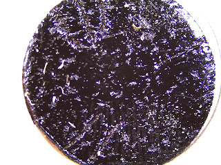
Between 2 & 3: Gypsophila
Between 3 & 1: Delphinium
Between 1 & 2: Alstroemeria
When preparing for the SEM we followed the standard procedure for safely putting our stub inside. Once the stub had been displayed on the screen we found individual pollen grains for each of our samples. At this point we photographed each grain and measured their widths. We then zoomed in to see the surface texture on each pollen grain.
Once this was done we had enough data to prove or disprove our hypothesis. When comparing the widths of the pollen grains to the widths of the flowers, we discovered that in our sample of Alstroemeria aurea, Gypsophila paniculata, and Delphinium parishii the pollen grain size was proportionate to the size of the flower.

Above is our stub photographed at 35x on the Leica microscope.
Between 2 & 3: Gypsophila
Between 3 & 1: Delphinium
Between 1 & 2: Alstroemeria
When preparing for the SEM we followed the standard procedure for safely putting our stub inside. Once the stub had been displayed on the screen we found individual pollen grains for each of our samples. At this point we photographed each grain and measured their widths. We then zoomed in to see the surface texture on each pollen grain.
Once this was done we had enough data to prove or disprove our hypothesis. When comparing the widths of the pollen grains to the widths of the flowers, we discovered that in our sample of Alstroemeria aurea, Gypsophila paniculata, and Delphinium parishii the pollen grain size was proportionate to the size of the flower.
No comments:
Post a Comment